Welcome to Test Slides, the second (*) online store of Diatom Lab! Our preparations are made for eternity and allow you to observe Diatoms and other microscopic objects in Very High Definition! Diatom Lab is the state-of-the-art scientific laboratory company founded in 2016 (VAT number IT 01635810193) that specializes in ground-breaking Micromanipulation services, Test slides for microscopy, Scientific photography / Microscopy imaging services.
“The use of Test Diatoms to check the quality of objectives is only one aspect of their use. The Test Diatoms should be also used to make sure that the whole system of the microscope is working well. That is the illumination as well as the combination of objective and eyepiece. It should become a habit to use a test slide every time the microscope is set up. In this way one ensures that what one sees is really there, not false and blurry images, and that one is missing nothing which should be visible with the optical system being used. Diatoms fill this requirement better than any other object” (from an old N.B.S. leaflet).
(*) For more microscope slides of Diatoms, Radiolarians and other microscopic objects, please visit the main site https://www.diatomshop.com/
Diatom Lab produces prepared microscope slides of the highest quality: our laboratory stands out for the use of unique cutting-edge materials, technologies and procedures. For further information please visit the pages "Innovative preparations" and "Diatom Lab. Clients and reputation".
We produce microscope slides for many important microscope manufacturing companies, government agencies, universities, laboratories, researchers and scientists, but also private individuals and amateurs are welcome: our materials and techniques are therefore not amateur and require cutting-edge, expensive instrumentation that goes beyond that used by a average, non-specialized laboratory. All our products are made in our internal laboratory. Our proprietary high refractive index mountants are not for sale as they require additional specific procedures and tools to be used.
We safely ship Worldwide: all parcels are fully tracked, so that you can check the progress of your items through to delivery! We have customers in many countries including the UK, USA, Europe, Asia, Africa and Oceania, and look forward to working with you! Combined shipping: worldwide shipping by registered international parcel is always 11,90 euros (with
the exception of the United States, where shipping via tracked
express parcel is possible at a cost of 40 euros), no matter how many items are in the cart! Payments are accepted with PayPal (no PayPal account needed, just a credit card!) or bank transfer. Diatom Lab works with honesty, accuracy and transparency: you'll always receive a proper invoice with every purchase.
Interesting facts about Diatoms: they are unicellular organisms, specifically microalgae, and live in waterways, oceans, and soils; they produce approximately 20 to 50 percent of the oxygen on Earth; they are primary producers in the food chain and constitute nearly half of the organic material found in the oceans; since the discovery of the microscope, architects and artists have been inspired by Diatom forms.
"Few objects are more beautiful than the minute siliceous cases of the Diatomaceæ: were these created that they might be examined and admired under the higher powers of the microscope?” (Charles Darwin, On the Origin of Species).
Click on the hamburger button at the top right to open the Menu.
DIATOM TEST SLIDES FOR OPTICAL MICROSCOPES (MICROSCOPE SLIDES OF SELECTED, MICROMANIPULATED TEST DIATOMS). TEST YOUR MICROSCOPE! Venture into the Micro and Nano structures! DIATOM TEST SLIDES ARE EXTREMELY USEFUL TO: A) evaluate and compare microscope models, objectives, eyepieces, condensers, light sources, optical filters, microscope cameras and microscope illumination techniques such as bright field, oblique illumination, differential interference contrast (DIC), phase contrast, dark field, polarized light, Rheinberg illumination, and Hoffman modulation contrast! B) quantify resolution in optical microscopes (defined as the smallest distance between two points on a specimen that can still be distinguished by the observer or camera system as two separate entities); C) practice using your microscopes at their highest levels! D) examine the variations in contrast and resolution by regulating the condenser aperture diaphragm; E) understand the importance of correction collar for minimazing spherical aberration.
Our diatom test slides are also purchased and used by several microscope manufacturing companies for quality control of their lenses and for demonstration purposes towards their customers!
All micromanipulated Diatoms are guaranteed to be fixed directly to the UNDERSIDE of the custom, optical quality cover glass (and not, as is common, on the microscope slide) for maximum resolution and contrast! (The reason for this: microscope objective performance drops quickly and noticeably as the specimen distance from the cover glass increases. Microscope objective lenses are designed to be optimally corrected for objects located immediately below the coverslip!)
- Barone, S. 2023. Diatoms: the best microscopic objects to check, set and compare optical microscopes and contrast techniques, Microscopy and Analysis, 65: 13-18 (ISSN 2049-4424) link
- Barone S., 2022. Diatom Lab Microscope Test Slide in commemoration of Edmund J. Spitta, Micscape Magazine, 321 (ISSN 1365 - 070x) link
David Walker is the editor of Micscape Magazine:
- Walker D., 2023. Exploring the Diatom Lab 'Microscope Test Slide in Commemoration of Edmund J. Spitta' with Near UV . Micscape Magazine, 328 (ISSN 1365 - 070x) link
- Walker D., 2021. Note on 'diatom dotting' at low magnifications. Resolving Stauroneis phoenicenteron, Micscape Magazine, 306 (ISSN 1365 – 070x) “Acknowledgement: The author used the invaluable 'Test Slide version 2.0' (Diatom Cubed mountant) supplied by Stefano Barone of Diatom Lab"
- Bulletin of the Quekett Microscopical Club, Nov 2019, No.77, ISSN 1350-9128, pp 22-25: the article recognizes Diatom Lab preparations as expertly-prepared and three microscope images of our slides have been published, such as a detail of the Diatom Test Slide version 2.0 with the comment “image chosen by editor to show the incredible resolution of this excellent slide”
Diatom Test Slide version 2.0 Click on the PDF button to download the free instructions (Updated version)  | Microscope Test Slide in commemoration of Edmund J. Spitta. Click on the PDF button to download the free instructions  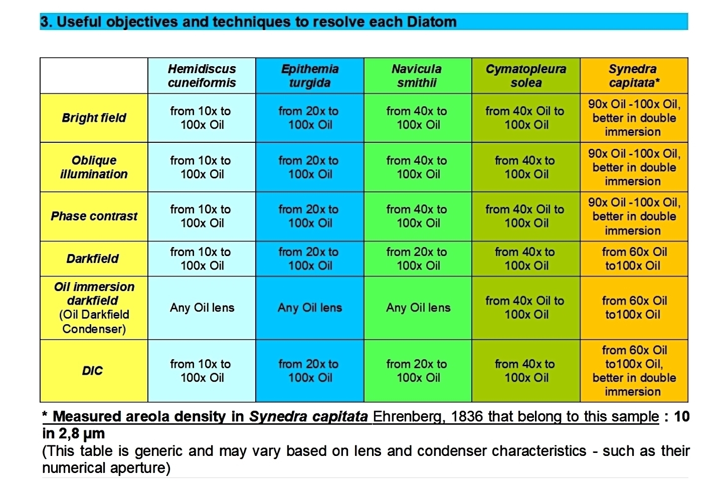 | Free article: Barone, S. 2023. Diatoms: the best microscopic objects to check, set and compare optical microscopes and contrast techniques, Microscopy and Analysis, 65: 13-18  |

Diatom Test Slide version 2.0. Download the free Instruction manual (Updated edition) by the website! The Diatom Test Slide version 2.0 groups Test Diatoms ranging from easy to extremely difficult. Since its appearance in 2018, hundreds of laboratories, microscope companies and microscopists from all over the world use this standardized slide with profit and satisfaction! NOT ALL Pinnularia nobilis (Ehrenberg) Ehrenberg ARE THE SAME! The MOST DIFFICULT Pinnularia nobilis (Ehrenberg) Ehrenberg sample among all the others we keep was chosen for the Diatom Test Slide version 2.0 (measurements were performed using a scanning electron microscope or SEM), in fact not all Pinnularia nobilis (Ehrenberg) Ehrenberg in the world have the same poroids measurements!
249.00 €
Add
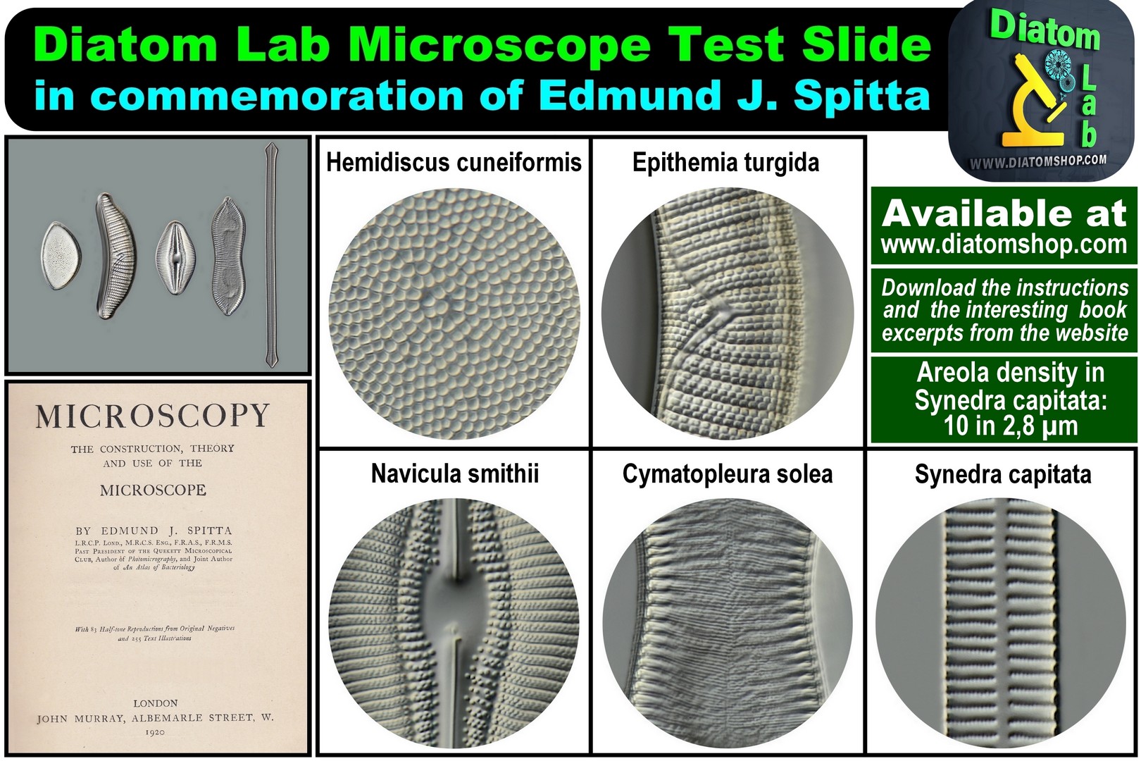
Diatom Lab Microscope Test Slide in commemoration of Edmund J. Spitta. A Complete Test for all dry and oil immersion lenses from 10x to 100x, in Bright Field, Oblique illumination, Phase contrast, Darkfield, Oil immersion darkfield and DIC. Download the instructions and the interesting book excerpts from www.diatomshop.com website
249.00 €
Add
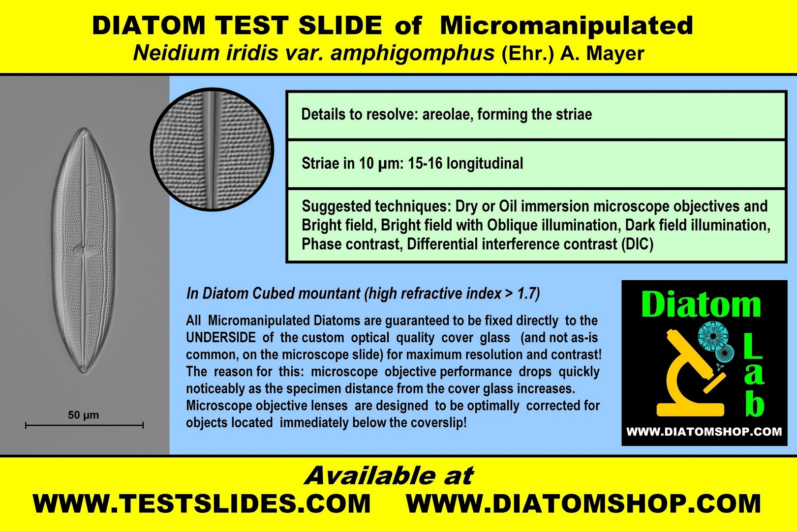
DIATOM TEST SLIDE of Micromanipulated Neidium iridis var. amphigomphus (Ehr.) A. Mayer
Details to resolve: areolae, forming the striae
Striae in 10 µm: 15-16 longitudinal
Suggested techniques: Dry or Oil immersion microscope objectives in Bright field, or Bright field with Oblique illumination, or Dark field illumination, or Phase contrast, or Differential interference contrast (DIC)
In Diatom Cubed mountant (refractive index > 1,7)
69.00 €
Add
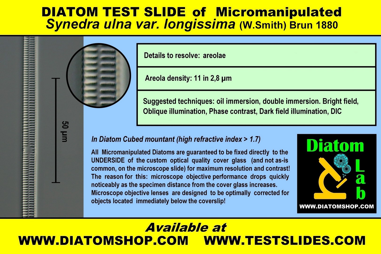
DIATOM TEST SLIDE of Micromanipulated Synedra ulna var. longissima (W.Smith) Brun 1880
Details to resolve: areolae
Areola density: 11 in 2,8 µm
Suggested techniques: Oil immersion, double immersion.
Bright field, Oblique illumination, Phase contrast, Dark field
illumination, DIC
Details to resolve: areolae
Areola density: 11 in 2,8 µm
Suggested techniques: Oil immersion, double immersion.
Bright field, Oblique illumination, Phase contrast, Dark field
illumination, DIC
69.00 €
Add

DIATOM TEST SLIDE of Micromanipulated Craticula cuspidata (Kützing) DG Mann ex Rotonda et al. (1990)
Details to resolve: very small areolae, forming the striae
Striae in 10 µm: 16-17 longitudinal
Suggested techniques: Oil immersion microscope objectives in Bright field, or Bright field with Oblique illumination, or Dark field illumination, or Phase contrast, or Differential interference contrast (DIC)
In Diatom Cubed mountant (refractive index > 1,7)
69.00 €
Add
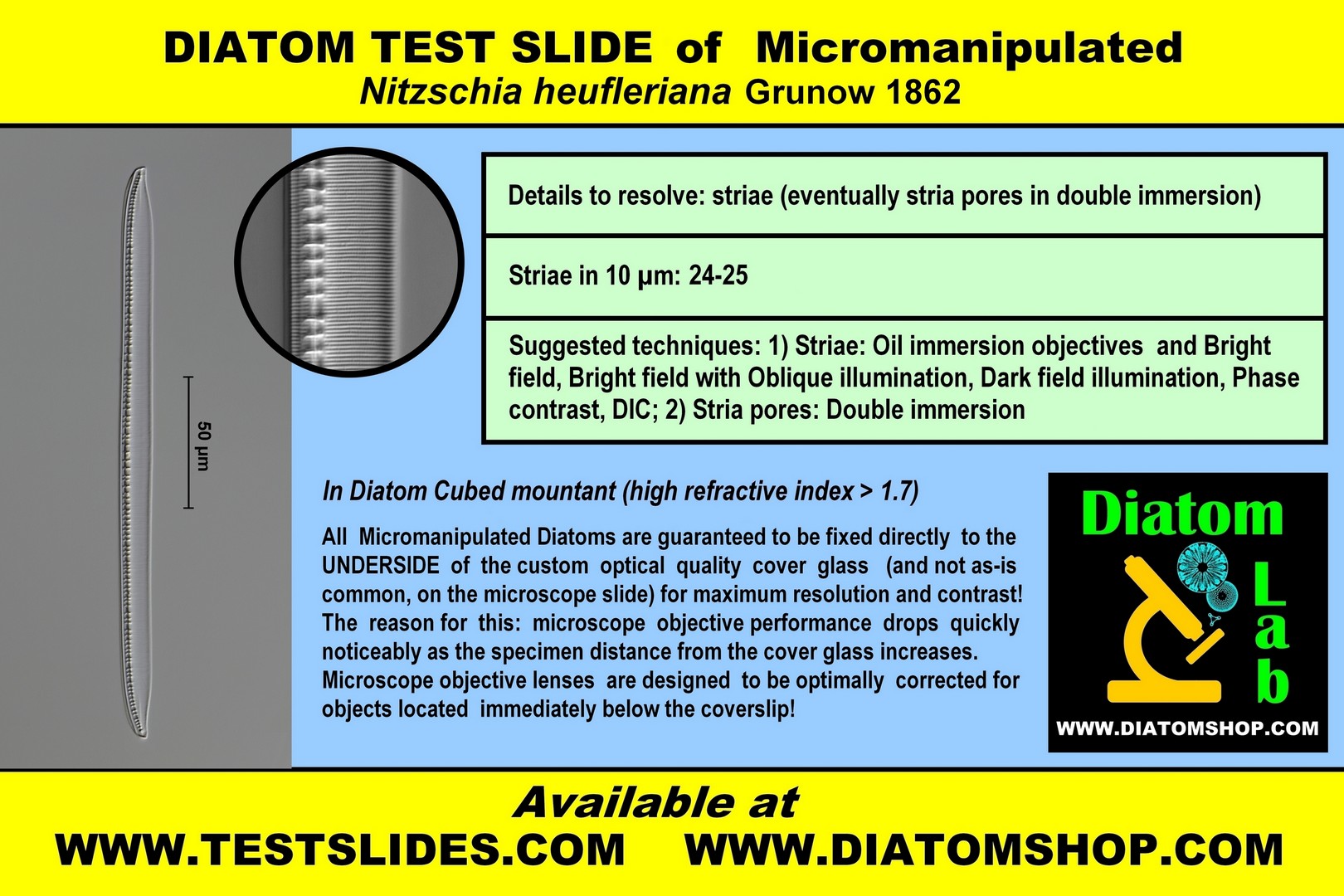
DIATOM TEST SLIDE of Micromanipulated Nitzschia heufleriana Grunow 1862
Details to resolve: striae or eventually stria pores in double immersion
Striae in 10 µm: 24-25
Suggested techniques:
1) Striae: Oil immersion microscope objectives in Bright field, or Bright field with Oblique illumination, or Dark field illumination, or Phase contrast, or Differential interference contrast (DIC);
2) Stria pores: Double immersion (= Oil immersion objective and Oil immersion condenser)
In Diatom Cubed mountant (refractive index > 1,7)
69.00 €
Add
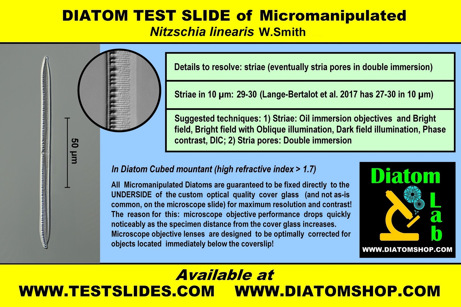
DIATOM TEST SLIDE of Micromanipulated Nitzschia linearis W.Smith
Details to resolve: striae or eventually stria pores in double immersion
Striae in 10 µm: 29-30, parallel
Suggested techniques:
1) Striae: Oil immersion microscope objectives in Bright field, or Bright field with Oblique illumination, or Dark field illumination, or Phase contrast, or Differential interference contrast (DIC);
2) Stria pores: Double immersion (= Oil immersion objective and Oil immersion condenser)
In Diatom Cubed mountant (refractive index > 1,7)
69.00 €
Add

DIATOM TEST SLIDE of Micromanipulated Pinnularia dactylus var. dariana (A.Schmidt) Cleve
Details to detect: poroids
The theoretical limit of resolution of most light microscopes is ∼ 0.2 μm, but these poroids can be detected by the techniques below, thanks to Diatom Cubed high refractive index mountant!
Viewed with SEM, QUANTITATIVE FEATURES in Diatom Lab's sample are:
Poroids inside the striae: 44 - 45 in 10 µm
Average distance between poroids: 0,072 - 0,089 µm. See SEM image from the same sample!
(While In Amphipleura pellucida striae number 37-45 in 10 µm and pores are spaced 0,16 - 0,19 µm apart)
Recommended microscope objectives: Oil-immersion 63 or 100x objectives having a very good or excellent numerical aperture (1,3 or 1,4);
Suggested techniques to detect the poroids: Double immersion (= Oil immersion objective and Oil immersion condenser) with Polarized light (the polarizers should be oriented perpendicular to each other = maximum level of extinction) and possibly oblique illumination; or Immersion dark field condenser (better 1,2/1,4) with Polarized light (the polarizers should be oriented perpendicular to each other = maximum level of extinction)
In Diatom Cubed mountant (refractive index > 1,7)
79.00 €
Add

DIATOM TEST SLIDE of Micromanipulated Surirella striatula Turpin (frustule has a strong torsion along the apical axis)
Details to detect: areolae, forming the striae
Viewed with SEM, the average distance between areolae is 0,14 µm.
The theoretical limit of resolution of most light microscopes is ∼ 0.2 μm, but these areolae can be detected by the techniques below, thanks to Diatom Cubed high refractive index mountant!
Recommended microscope objectives: oil-immersion 63 or 100x objectives having a very good or excellent numerical aperture (1,3 or 1,4)
Suggested techniques: Double immersion (= Oil immersion objective and Oil immersion condenser) in: Polarized light (the polarizers should be oriented perpendicular to each other = maximum level of extinction) with possibly oblique illumination; or Immersion dark field condenser (better 1,2/1,4) with Polarized light (the polarizers should be oriented perpendicular to each other = maximum level of extinction)
In Diatom Cubed mountant (refractive index > 1,7)
69.00 €
Add
TEST INTEGRATED CIRCUITS for light microscopes in reflected light, stereo microscopes, scanning electron microscopes (SEMs), objectives for macro photography

Very small integrated circuit (0.75mm X 0.66mm) with many fine details, dry mounted on microscope slide without cover glass. Excellent for testing light microscopes in reflected light, stereo microscopes, scanning electron microscopes (SEMs), objectives for macro photography! This test integrated circuit has been discussed in the following article: Walker D., Exploring the Diatom Lab prepared slide of an integrated circuit chip, Micscape Magazine, April 2020, Issue 289, ISSN 1365 – 070x: "The slide is impeccably prepared / mounted", "In my view the slide is excellent value for money and maintains the high standards set by the Diatom Lab shop (I have a number of the diatom slides)"
69.00 €
Add
OPTICAL ABERRATIONS DETECTOR. It's a very useful Diatom Lab microscope slide that detects optical aberrations - even the most imperceptible ones - in light microscopy, such as: a) Curvature of field (the image appears sharp and crisp in the center or on the edges of the viewfield but not both); b) Distortion (changes in the shape of the image); c) Barrel distortion and Pincushion distortion; d) Spherical aberration (the specimen image appears hazy or blurred and slightly out of focus); e) Chromatic aberration (presence of color fringes at the edges of the borders); f) Astigmatism (the ideal circular point image blurs into a diffuse circle, elliptical patch, or line). Thanks to this unique microscope slide, you can easily test and compare your objectives, eyepieces, condensers, phototubes, microscope cameras, and magnification changers! This is an amazing microscope slide in the center of which there are, incredibly, 90.000 microscopic squares having a side of 10 µm (ten micrometres or microns) and 10 µm apart! These microscopic squares also form nine larger squares useful in case of low magnification (see the page "Optical Aberrations Detector, Centering Device, Micrometers" for better image quality). This slide is excellent for testing macro lenses as well! Click on this link to download the pdf file. All Rights Reserved.
Optical Aberrations Detector for transmitted light (with cover glass and mountant).
249.00 €
Add
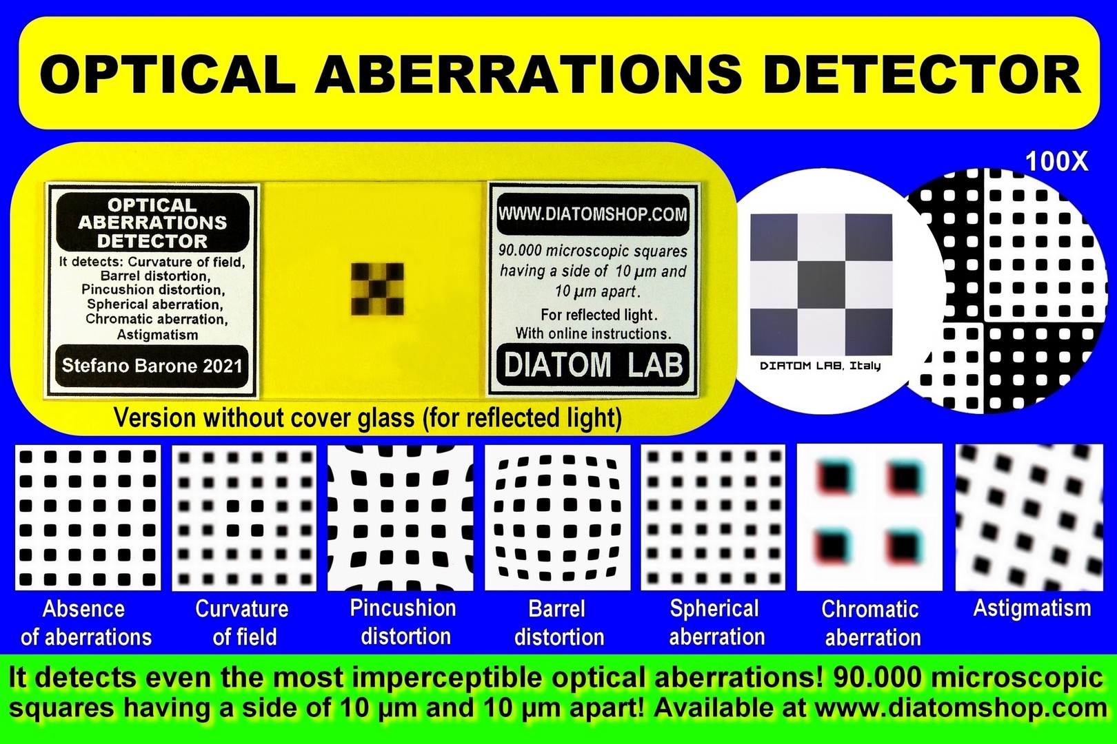
Optical Aberrations Detector for reflected light (without cover glass).
249.00 €
Add
CENTERING DEVICE & MICROMETER GLASS SLIDE. It's an all-in-one microscope slide that incorporates a centering device to center the microscope and the camera and a stage micrometer for microscope calibration. The centering device consists of 25 perfect concentric circles, 50 µm (fifty micrometres or microns) thick and 50 µm apart! It is very useful to center the microscope parts (condenser top lens, field iris diaphragm, prisms of the binocular/trinocular head, etc.) and the microscope camera. (It is advisable to make changes to the microscope only if you are an expert.) The stage micrometer for microscope calibration is a 1 mm Scale with 0,01 mm (10 µm) divisions; the slide can be used in combination with a micrometer eyepiece or a measurement software (for cameras) in order to measure preparations. (See the page "Optical Aberrations Detector, Centering Device, Micrometers" for better image quality.) All Rights Reserved.
For transmitted light (with cover glass and mountant). Diatom Lab CENTERING DEVICE & MICROMETER GLASS SLIDE is an all-in-one microscope slide that incorporates a centering device to center the microscope and the camera and a stage micrometer for microscope calibration. The centering device consists of 25 perfect concentric circles, 50 µm (fifty micrometres or microns) thick and 50 µm apart! It is very useful to center the microscope parts (condenser top lens, field iris diaphragm, prisms of the binocular/trinocular head, etc.) and the microscope camera. The stage micrometer for microscope calibration is a 1 mm Scale with 0,01 mm (10 µm) divisions. The slide can be used in combination with a micrometer eyepiece or a measurement software (for cameras) in order to measure preparations.
This product is unavailable.
99.00 €
Add
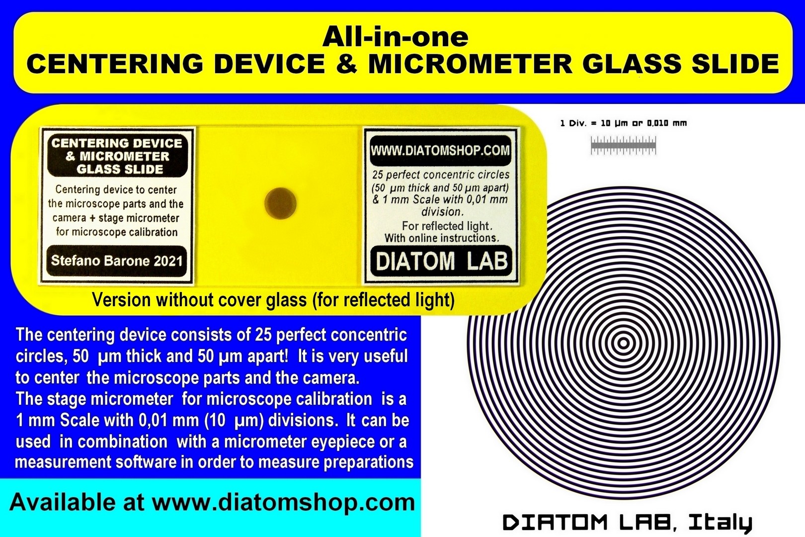
For reflected light (without cover glass). Diatom Lab CENTERING DEVICE & MICROMETER GLASS SLIDE is an all-in-one microscope slide that incorporates a centering device to center the microscope and the camera and a stage micrometer for microscope calibration. The centering device consists of 25 perfect concentric circles, 50 µm (fifty micrometres or microns) thick and 50 µm apart! It is very useful to center the microscope parts (condenser top lens, field iris diaphragm, prisms of the binocular/trinocular head, etc.) and the microscope camera. The stage micrometer for microscope calibration is a 1 mm Scale with 0,01 mm (10 µm) divisions. The slide can be used in combination with a micrometer eyepiece or a measurement software (for cameras) in order to measure preparations.
This product is unavailable.
99.00 €
Add
CUSTOMIZED ARRANGEMENTS, SERVICES
Specifically mounted diatom frustules for photonic researches and applications. In fact Diatoms are photonic crystals (photonics is the skill of manipulating photons): they exhibit sophisticated optical systems. Diatom Lab has collaborated with various institutes by providing specifically mounted diatom frustules for many researches. Price: ask for a free quote
This product is unavailable.
0.00 €
High-resolution microscope images and microscope videos (exclusive or non-exclusive licences): Diatom Lab has won several international scientific photography awards. We provide scientific imaging services using our state of the art Zeiss Axio Imager.A2 and other microscopes in full frame camera format. Various illumination techniques are available. Price: ask for a free quote
This product is unavailable.
0.00 €
New publications, articles for magazines: ask for a free quote
This product is unavailable.
0.00 €
Please feel free to contact us with questions regarding quotes for customized products and services. We will do our best to answer your request as soon as possible.
Diatom Lab is based in Europe (Italy), customers are responsible for paying any assessed customs fees upon receipt. We are not responsible for any customs fees charged. We reserve the right to make changes to our products without prior notice. Since these products are partially hand-made in our laboratory, the products you receive may be different from those pictured here. Return
policy: the buyer is responsible for the cost of shipping the item
back to the seller. A refund will be issued once the item is returned
to the sender in the same condition as it was originally sold.
Customers have 14 days from the date they receive their order to
notify the seller of their intention to return an item, and another
14 days to actually return it.
For more microscope slides of Diatoms, Radiolarians and other microscopic objects, please visit the main site https://www.diatomshop.com/
"The revelations of the Microscope are perhaps not excelled in importance by those of telescope. While exciting our curiosity, our wonder and admiration, they have proved of infinite service in advancing our knowledge of things around us" (Joseph Leidy)